Figure 2.

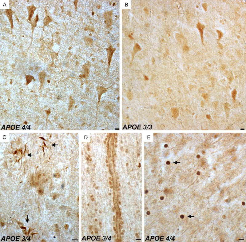
Detection of fragmented apoE in the frontal cortex of the Alzheimer’s disease brain. Representation bright-field staining in AD frontal cortex tissue sections following application of the nApoeCFp17 antibody (1:100). (A and B) Representative immunostaining of neurons by nApoECFp17 in two separate AD cases that differ in the APOE allele genotype as depicted in (A and B). Staining was also observed in neuropil threads within plaque-rich regions (arrows, C), along blood vessels (D), and the strongest labeling was within small circular structures throughout both gray and white matter (arrows, E). All scale bars represent 10 µm.
