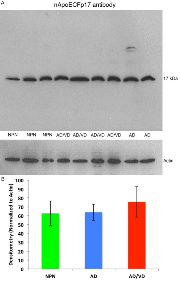
Figure 4.

Western blot analysis of human brain extracts with the nApoECFp17 antibody. (A) Western blot analysis was performed utilizing protein extracts prepared from frozen, frontal cortex brain tissue from neuropathological controls, (NPN, APOE genotype 3/4), AD/VD subjects (APOE genotype 4/4), or AD subjects (APOE genotype 3/4). Soluble, human brain lysates were separated on 15% SDS-PAGE gels, transferred to nitrocellulose, and then probed with the nApoECFp17 antibody (1:500). (A) A prominent band at the predicted molecular weight for the nApoE1-151 fragment was observed in all cases examined. The lower panel in (A) represents a loading control blot using a rabbit antibody to beta-actin (1:400). (B) Densitometry analysis of the p17 band normalized to actin in NPN subjects, AD, and AD/VD subjects indicated no significant differences between any of the cohorts tested. Data represent the combined densitometry average for two independent experiments, ± S.D.
