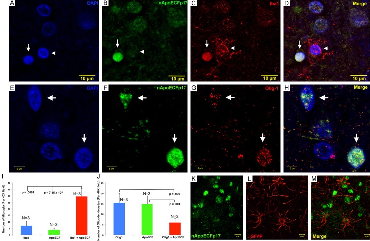
Figure 5.
Nuclear localization of an amino-terminal fragment of apoE within microglia of the AD brain. (A-D) Representative images from confocal immunofluorescence in AD utilizing the nuclear stain, DAPI, (A), nApoECFp17 (B), the microglial specific marker, Iba1 (C), with the overlap image shown in (D). Note the strong nuclear localization of the nApoECFp17 antibody (arrow) within labeled microglia. The arrowhead in (A-D) depicts a single microglial cell labeled with Iba1 that was not strongly labeled with the nApoECFp17 antibody. (E-H) Identical to (A-D) with the exception that the oligodendrocyte marker, Olig-1, was employed to specifically label oligodendrocytes. (I, J) Quantification of microglia (I) or oligodendrocytes (J) double-labeled with nApoECFp17 indicated co-localization in 80% and 27.5% of microglia and oligodendrocytes, respectively. Data show the number of Iba1- or olig-1-labeled cells alone (blue bars), number of nApoECF-labeled nuclei (green bars), and the number of cells with both antibodies (red bars) identified in a 40× field within frontal cortex AD sections by immunofluorescence microscopy (n=3 fields for 3 different AD cases, ± S.E.M.). (K-M) In contrast to the nuclear localization of nApoECFp17 within microglia and oligodendrocytes, confocal immunofluorescence double-label experiments with nApoECFp17 and the astrocytic marker, GFAP, failed to produce any co-localization between these two markers.

