Figure 6.

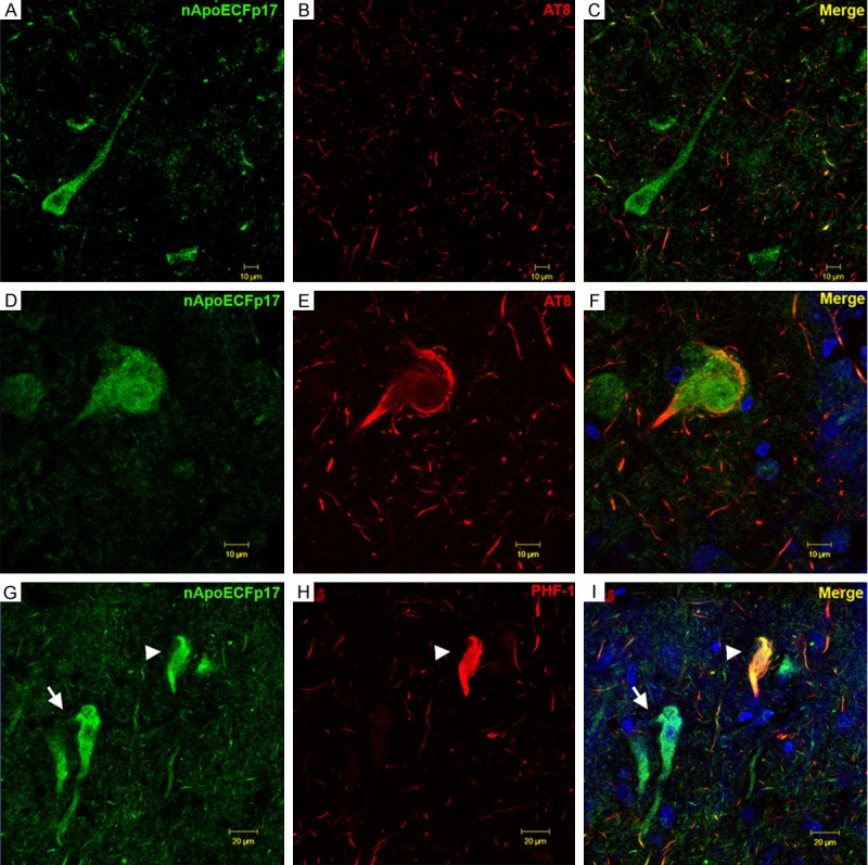
Co-localization of a fragment of apoE within a subset of neurofibrillary tangles in the AD brain. (A-F) Confocal immunofluorescence double-labeling in representative frontal cortex sections of the AD brain utilizing the nApoECFp17 antibody (green) and AT8 (red) revealed co-localization of the two antibodies in a subset of neurons (F), but not in the majority of neurons labeled with nApoECFp17 (C). Note that even though the two antibodies localized to the same NFT, there appeared to be a spatial separation with AT8 labeling staining the periphery and nApoECFp17 staining the interior of the cell (F). (G, H) Identical to (A-F) with the exception that the mature tangle marker, PHF-1 (red, H) was employed. Similar results with PHF-1 were observed with only a subset of fibrillar NFTs showing co-localization of the two antibodies (arrowhead, I). In contrast to AT8 labeling, strong co-localization of PHF-1 and nApoECFp17 was evident within the NFT (arrowhead, I). The blue fluorescence in (F and I) represents the nuclear stain, DAPI.
