
Figure 4.
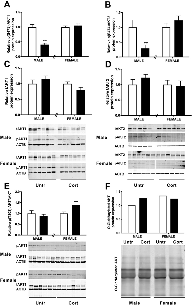
The effects of corticosterone exposure (black bars) on (A) serine 473 phospho-AKT1, (B) serine 474 phospho-AKT2 along with (C) total AKT1 and (D) total AKT2 protein in whole placentae of male and female C57/BL6 mouse fetuses at E14.5 compared with untreated controls (white bars). (E) Phosphorylation of AKT1 on threonine 308 relative to total AKT1 was also examined. Pooled male and female E14.5 placental tissue (n = 6) was further immunoprecipitated with RL2 antiserum to isolate O-GlcNAcylated proteins and immunoprobed with pan Akt antiserum following SDS-PAGE (F). Data represents mean + SEM, **P < 0.01. Protein expression examined by western blot with β-Actin (ACTB) as loading control, normalised to untreated control of the same sex (Top band analysed for tAKT and both bands analysed for O-GlcNAcylated AKT). Analysed using a two-tailed t-test with untreated controls of the same sex (n = 6).