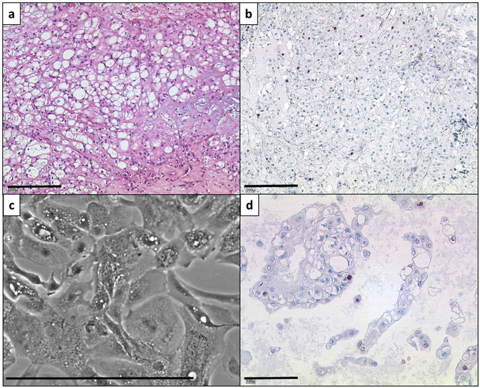
Figure 1.

Morphology and proliferation rate detection of the parental tumour and the U-CH14 cell line. (a) Section of the parental tumour. The cells show the typical physaliphorous appearance. The tumour is classified as not otherwise specified (NOS) subtype. (b) Ki-67 staining of a parental tumour section. The proliferation index is below 5%. (c) Phase contrast image of the U-CH14 cell line. The typical morphology is conserved in vitro. (d) Proliferation index of U-CH14 (<5%; Ki-67 staining). Lines indicate 200 µm (a+b) or 100 µm (c+d).
