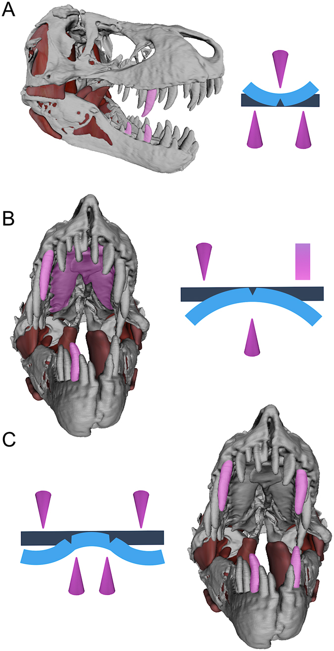
Figure 4.

Jaw models of Tyrannosaurus rex paired with idealized beam diagrams, illustrating three- (A) (lateral view), (B) (anterior view) and four-point ((C), anterior view) loading configurations that allowed T. rex to promote failure stresses and fracture rigid structures (e.g., bone) without the aid of occluding dentitions. Teeth (cones) and the osseus palate, composed of the right and left maxillae and an anterior expansion of the vomer (rectangle), are shown as contact points in pink; original beam shapes are dark blue; and idealized plastic deformations (exaggerated) are light blue.
