
Figure 2.
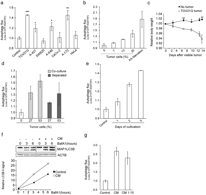
TOV21G cancer cells cause cachexia and secrete autophagy-inducing substances. (a) Screen of various cancer cell lines for the ability to induce autophagy in reporter cells (co-cultures). Mean from one experiment using triplicate wells ± SD. *p = 0.04, **p = 0.02, ***p = 0.008 vs control (Student t-test). (b) Reporter cells co-cultured with increasing percentage of TOV21G cells. Amino acid starvation (HBSS, 16 h) as positive control. Mean from one experiment representative of six experiments using duplicate wells ± SD. (c) Mean relative body weight (at indicated time intervals) of TOV21G tumor-bearing mice (n = 6) and control mice (n = 6) ± SEM. *p < 0.01, **p < 0.005 vs no tumor (Student t-test). (d) Autophagy flux in reporter cells co-cultured with or separated from TOV21G cells using a semi-permeable membrane. Mean from one experiment using duplicate wells ± SD. (e) Autophagy flux in reporter cells after 3 days of exposure to conditioned medium (CM) from TOV21G cells cultured for 1–3 days (n = 1 for 1 day and n > 6 for 3 days). Mean from one representative experiment using duplicate wells ± SD. (f) Protein levels of MAP1LC3B in reporter cells treated with CM from TOV21G cells for 3 days with or without BafA1 (100 nM) for indicated time periods. MAPLC3B-II signal is normalized against β-actin/ACTB. The data are representative for two independent experiments. Image, originating from the same blot, is cropped. (g) Autophagy flux in reporter cells exposed to diluted (1:15 in complete growth medium) or undiluted CM from TOV21G cells. Mean from one representative of three independent experiments using triplicate wells ± SD.