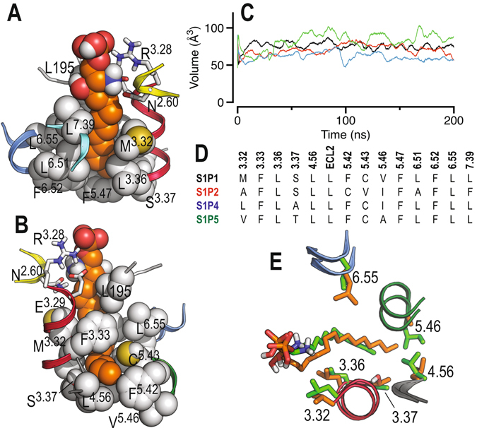
Figure 5.

Molecular dynamics simulations of sphingosine-1-phosphate receptors. (A) Detailed view of the narrow channel of S1P1 where the alkyl tail of S1P (in orange) must expand. The end of the channel is delimited by the amino acids at positions 4.56, 5.42 and 5.46. The structure depicts the final conformation at 0.2 μs. (B) Same as in panel A but rotated 180°. (C) Volume of the channel in S1P1 (black), S1P2 (red), S1P4 (blue) and S1P5 (green) along the MD trajectories with the natural S1P agonist as calculated with POVME49. (D) Sequence alignment, among sphingosine receptors, of the amino acids forming this channel. (E) The final conformation, at 0.2 μs, obtained in the MD simulations of S1P4 in complex with C16 (in green) and S1P5 in complex with C20 (in orange).
