
Figure 1.
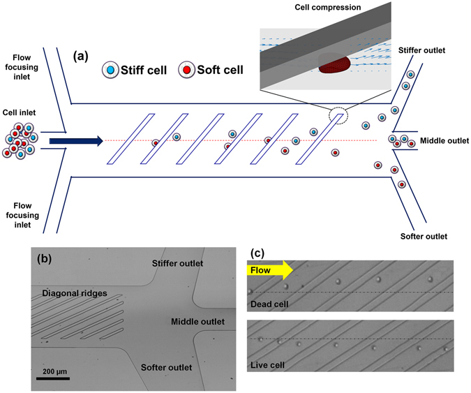
Ridge based microfluidic device for viable cell sorting. (a) Schematic representation of the device showing that cells will squeeze through ridges and flow towards different outlets based on their stiffness differences; (b) optical micrograph of the fabricated device showing the ridges and outlets; (c) representative micrograph of overlaid video of dead and live cells moving towards opposite directions in the device. The yellow arrow show the direction of fluid flow.