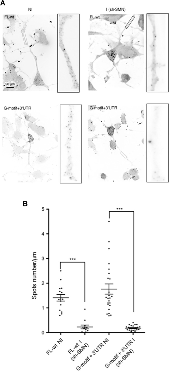
FIGURE 7.

Localization of the FL-wt and G-motif + 3′UTR reporter mRNAs in axons of differentiated NSC-34 cells is SMN-dependent. (A) Spots corresponding to the indicated mRNAs in axons of noninduced (NI) or induced (I) shSMN-NSC-34 cells are shown. Representative images of FISH experiments show the RNA spots localized in axons before and following SMN depletion. Scale bar, 20 µm. Insets on the right of each panel are enlarged images of boxed sections. (B) Quantitative analysis of localization of reporter RNAs in axons as illustrated in A. Scatter plots show the number of mRNA spots localized in the distal segment of axons with the FL-wt construct before (NI, noninduced, n = 19) and after depletion of SMN (I, induced; sh-SMN, n = 19, P = 0;001), and with the G-motif + 3′UTR reporter (NI, n = 24; I, n = 24, P < 0.0001). Mean and SEM are shown. Statistical significance was calculated using the Wilcoxon test.
