Figure 5.

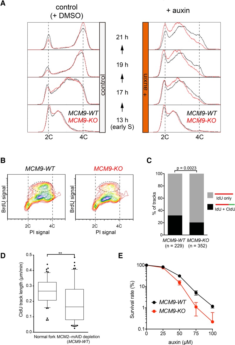
MCM8–9 promotes DNA synthesis after fork stalling. (A) MCM9-WT and MCM9-KO derivatives of the MCM2-mAID cells (shown with black and red lines, respectively) were synchronized and treated as in Figure 2. DNA content was then measured using flow cytometry after either DMSO (left) or auxin (right) treatment. (B) Incorporation of BrdU after fork stalling was examined using flow cytometry in the MCM9-WT and MCM9-KO backgrounds. (C) DNA synthesis after fork stalling was examined using isolated DNA fibers. The MCM9-WT and MCM9-KO derivatives of the MCM2-mAID cells were synchronized and treated as in Figure 2. At the 21-h time point, the cells were labeled with iododeoxyuridine (IdU) followed by chlorodeoxyuridine (CldU) labeling. The percentage of DNA fibers labeled with both IdU and CldU was compared with those that contained IdU only. n = 229 IdU; n = 352 CldU. (D) CldU tract length was quantified in the wild-type (normal fork) and MCM2-mAID cells with MCM9-WT. (E) Cells with a reduced amount of MCM2–7 are more reliant on MCM8–9 for their survival. MCM9-WT and MCM9-KO derivatives of the MCM2-mAID cells (shown in black and red lines, respectively) were allowed to form colonies in the presence of various doses of auxin. The colony number was counted after crystal violet staining. The survival rate in the absence of auxin was defined as 100%.
