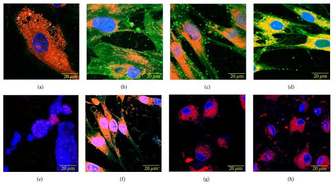
Figure 6.
Cytotoxic effect on cultured dental pulp stem cells (DPSCs) exposed to 50 μmoL L−1 CatDex-FITC and CHX for different amounts of time. (a) Negative control; (e) positive control. (b), (c), and (d): CatDex-FITC at 3 sec, 1 h, and 5 h, respectively. (f), (g), and (h): chlorhexidine at 3 sec, 1 h, and 5 h, respectively. In red, emission of MitoTracker-H2XRos staining mitochondria; in blue, DAPI signal localised to the nuclear compartment; in green, CatDex-FITC signal localised to cytoplasm; and in orange, colocalisation of MitoTracker and FITC signals. Confocal laser microscope, fluorescent, histochemical technique, and objective 63x.

