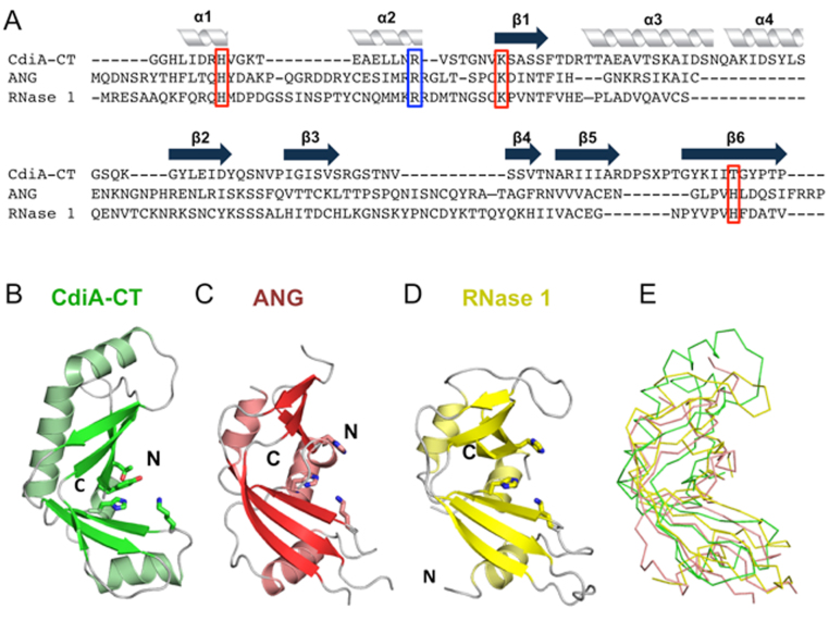
Figure 2.
The CdiA-CTYkris nuclease domain adopts the RNase A fold. (A) Structure-based alignment of the CdiA-CTYkris nuclease domain with human angiogenin (ANG) and mouse RNase 1. Secondary structure elements are indicated above the alignment. Putative catalytic and substrate-binding residues are boxed in red and blue, respectively. (B) CdiA-CTYkris nuclease domain. (C) Human ANG (PDB ID: 4B36 (35)). (D) Mouse pancreatic RNase 1 (PDB ID: 3TSR (36)). Structures in panels B–D are shown in cartoon representation with catalytic residues depicted in stick representation. (E) Superimposition of CdiA-CTYkris with ANG and RNase 1 in ribbon representation.

