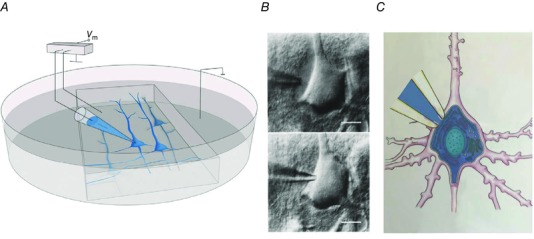
Figure 1. Whole‐cell voltage recording from a pyramidal cell in a parasagittal brain slice.

A, brain slice mounted in an experimental chamber for whole‐cell voltage recording via a patch pipette. B, differential interference contrast images of pipette tip approaching the cell body of a neuron and ‘cleaning’ of extracellular neuropil by positive pressure applied to the pipette solution (upper photomicrograph). Termination of positive pressure causes collapse of glial and neuropil tissue around the pipette tip and sealing of the pipette tip to the cell membrane (lower photomicrograph). Application of a brief pulse of negative pressure causes high‐resistance (gigaohm) contact between the pipette tip and cell membrane. The membrane patch drawn into the pipette tip is removed, e.g. by a brief increase in negative pressure (Hamill et al. 1981). Calibration bars represent 10 μm C, neuron cell body, proximal dendrites and initial segment of the axon. A pipette tip with open access between the pipette filling solution and the intracellular space of the neuron allows voltage recording and dialysing of the cell with pipette solution (blue).
