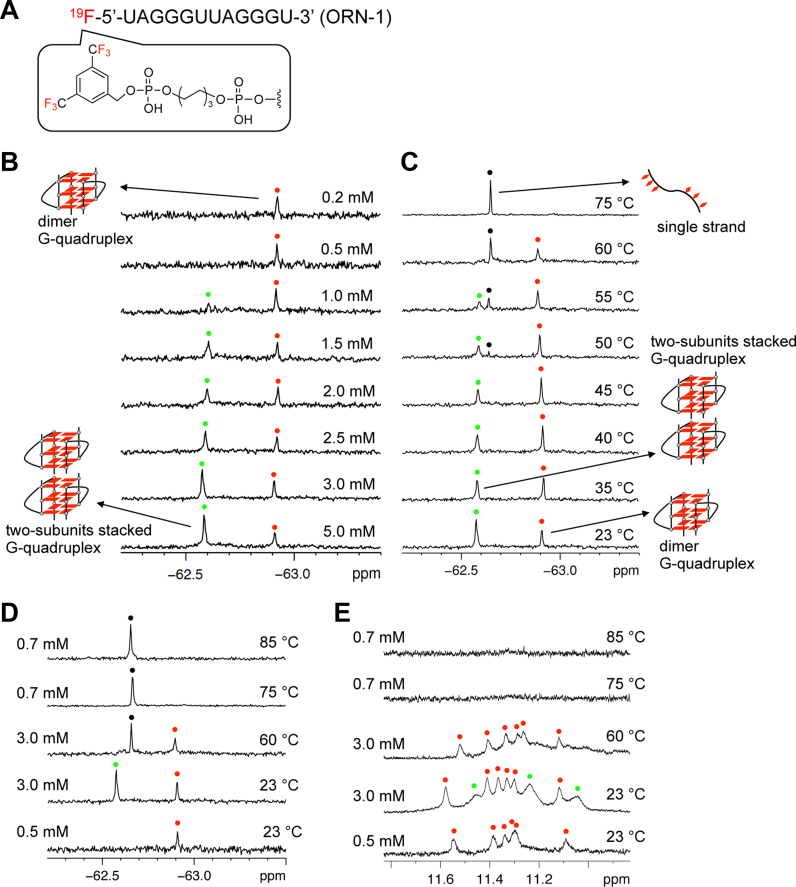
Figure 2.
19F NMR and 1H NMR spectra of 19F labeled RNA. (A) Chemical structure of 19F labeled telomere RNA bearing 3,5 bis(trifluoromethyl)phenyl group at the 5΄ terminal. (B) 19F NMR spectra of 19F labeled telomere RNA at different concentrations. The peaks of the dimeric and two-subunits stacked G-quadruplex are marked with red and green spots, respectively. Concentrations of RNA indicated on the right. (C)19F NMR of 19F labeled RNA at different temperatures. Red and green spots indicated dimer and two-subunits stacked G-quadruplex. The peaks of single strand are marked with black spots. Temperatures indicated on the right. Condition: 3 mM RNA in 50 mM KCl and 10 mM Tris-HCl buffer (pH 7.0). The sample is kept for 10 min at each temperature. (D) 19F NMR of 19F labeled RNA at different temperatures and concentrations. The peaks of dimer G-quadruplex are red spots. The peak of two-subunits stacked G-quadruplex is green spot. The peaks of single strand are marked with black spots. (E) 1H imino proton NMR of 19F labeled RNA corresponding to 19F NMR at different temperatures and concentrations. The peaks characteristic of the dimeric and two-subunits stacked G-quadruplex are marked with red and green spots, respectively.

