Figure 5.
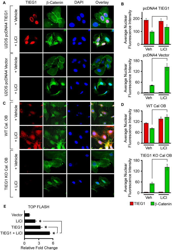
Effect of LiCl on β-catenin nuclear localization and activation of the TOP FLASH reporter in the presence and absence of TIEG1 expression. (A) U2OS cells were transfected with an empty vector (pcDNA4) or a TIEG1 expression vector and subsequently treated with vehicle control or LiCl for 24 h. Over-expressed TIEG1 protein (red) and endogenous β-catenin protein (green) were detected by immunofluorescence. DAPI staining (blue) indicates nuclei. (B) Quantitation of nuclear TIEG1 and β-catenin protein in U2OS cells transfected with an empty vector or TIEG1 expression construct. (C) Confocal microscopy images depicting endogenous levels of TIEG1 protein (red) and β-catenin protein (green) in WT and TIEG1 KO calvarial osteoblasts. (D) Quantitation of nuclear TIEG1 and β-catenin protein in WT and TIEG1 KO calvarial osteoblasts. (E) TOP FLASH reporter activity in U2OS cells that were co-transfected with empty vector (pcDNA4) or a TIEG1 expression vector and treated with or without LiCl as indicated. * denotes significance at P < 0.05 relative to empty vector controls.

