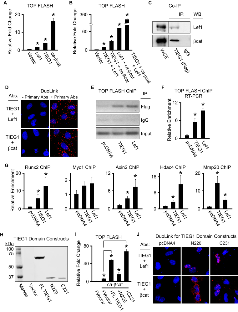
Figure 6.
TIEG1 interacts with and serves as a co-activator for Lef1 and β-catenin. (A and B) TOP FLASH reporter construct activity in U2OS cells transfected with empty vector or TIEG1, Lef1 and constitutively active β-catenin (ca-β-cat) expression vectors as indicated. (C) Co-immunoprecipitation assay in U2OS cells co-transfected with a flag-tagged TIEG1 expression construct and a Lef1 or β-catenin expression construct. Non-immunoprecipitated whole cell extracts (WCE) are shown as a loading control. (D) Duolink assays depicting interaction of TIEG1 with Lef1 and β-catenin (red fluorescent spots) in transiently transfected U2OS cells. Assays in which the primary antibodies targeting TIEG1, Lef1 and β-catenin were excluded are shown as negative controls. (E and F) Transient chromatin immunoprecipitation (ChIP) assays in U2OS cells indicating TIEG1 and Lef1 association with Tcf/Lef elements in the TOP FLASH reporter construct by both semi-quantitative (E) and real-time PCR (F). (G). ChIP assays in U2OS cells transfected with TIEG1 or Lef1 expression vectors demonstrating enrichment of TIEG1 and Lef1 on known Tcf/Lef enhancer elements encoded within endogenous canonical Wnt target genes. All ChIP data were normalized using input samples. (H) Western blot indicating protein expression levels of full-length (FL) TIEG1 or N-terminal (N220) and C-terminal (C231) domains of TIEG1 following transfection into U2OS cells. (I) TOP FLASH reporter construct activity in U2OS cells transfected with a constitutively active β-catenin expression vector (ca-βcat) and indicated TIEG1 expression vectors. * denotes significance at P < 0.05 relative to empty vector controls. (J). Duolink assays depicting interaction of the N- and C-terminal domains of TIEG1 with Lef1 and β-catenin (red fluorescent spots) in transiently transfected U2OS cells. Empty vector (pcDNA4) transfected cells are shown as negative controls.

