Fig. 1.

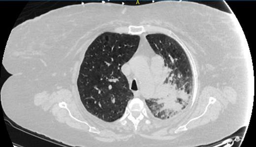
Chest computed tomography image showing a left upper lung mass associated with obstructive pneumonitis involving much of the left upper lobe.

Chest computed tomography image showing a left upper lung mass associated with obstructive pneumonitis involving much of the left upper lobe.