Fig. 2.

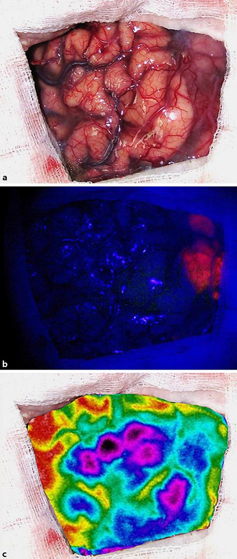
Intraoperative view of the patient's cerebral surface with the naked eye (a), under blue light exposure (b), and by using IRT brain mapping (c).

Intraoperative view of the patient's cerebral surface with the naked eye (a), under blue light exposure (b), and by using IRT brain mapping (c).