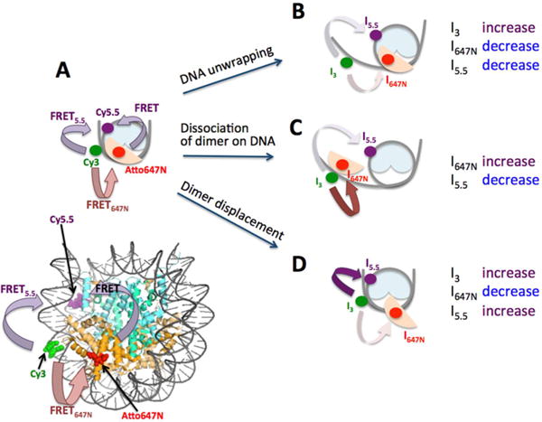
Figure 1.

Three-color FRET experimental setup to monitor the early steps of histone H2A-H2B dimer dissociation. (A) A schematic representation of a complete nucleosome core particle with intact DNA and H2A-H2B dimer reporting moderately high FRET5.5 and FRET647N. Fluorescence intensities from Cy3, Atto647N, and Cy5.5 are denoted by I3, I647N, and I5.5, respectively. (B) A nucleosome with unwrapped DNA and intact dimer would report decreasing I5.5 and I647N and increasing I3. (C) A nucleosome with DNA and dimer dissociating simultaneously would show decreasing I5.5 and increasing I647N. This mode of dissociation can be via gyre opening or DNA unwrapping, which cannot be distinguished in our setup. (D) A nucleosome with displaced dimer with intact DNA would report increasing I5.5 and I3, and decreasing I647N.
