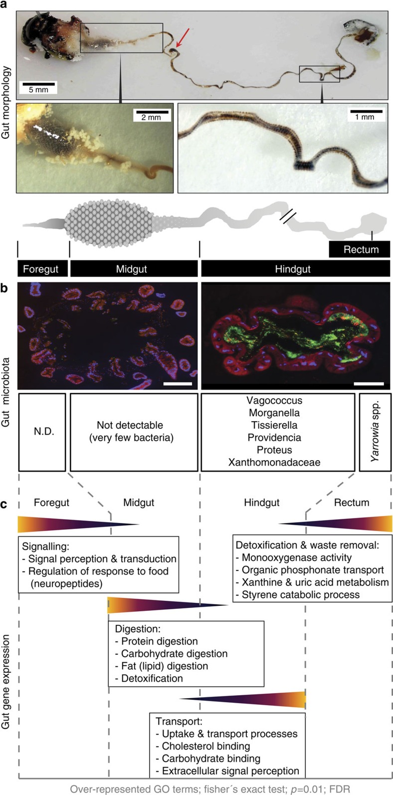
Figure 1. Gut transcriptome and gut microbial community of N. vespilloides.
(a) Morphology of a male N. vespillloides gut showing the complete gut structure as well as midgut and flat hindgut region with bulbous inflations. The red arrow indicates the position where the gut was separated into the midgut and hindgut portions during dissection. (b) Fluorescence in situ hybridization revealed some sections of the midgut with low (not detectable) bacterial prevalence, but a bacterially abundant hindgut region (bacteria stained with the general bacterial probe (green) EUB338-Cy5). Scale bars, 200 μm. (c) Differential representation of GO terms, reflecting metabolic differences between gut regions, with the foregut and midgut involved in signalling and digestion, the hindgut involved in the transport of nutrients and the rectum involved in waste removal and detoxification.

