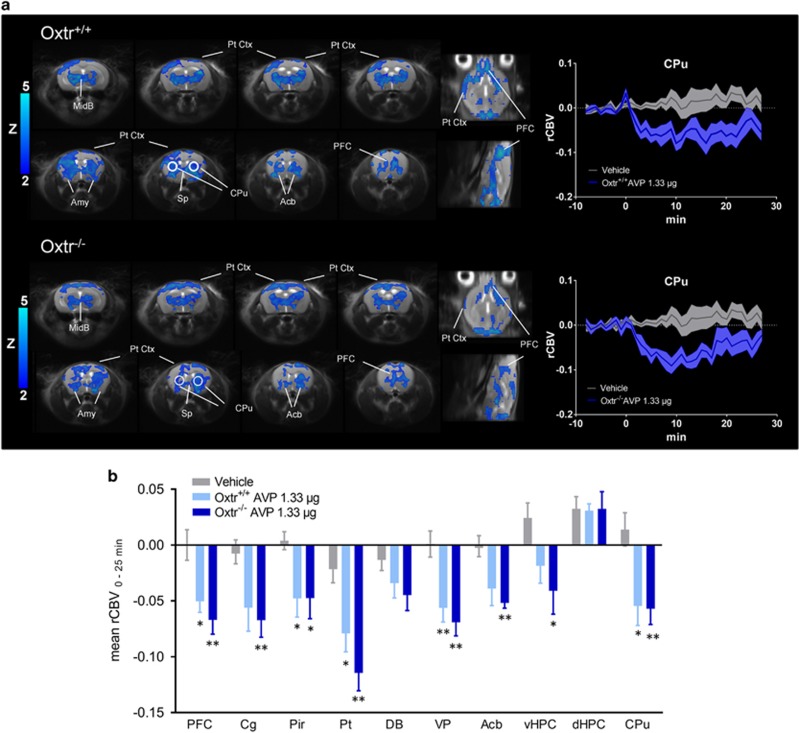
Figure 4.
fMRI deactivation produced by acute intranasal administration of AVP in the mouse brain. (a) AVP-induced fMRI response was mapped both in control Oxtr+/+ and Oxtr−/− mice (1.33 μg/mouse). The fMRI response was mapped with respect to vehicle-treated baseline (water) using a boxcar function, as we did not observe apparent regional differences in temporal fMRI dynamics (Supplementary Figure S5). An illustrative fMRI time course in a representative region of interest (identified by a circle in the activation maps) is reported. AVP or vehicle were administered at time 0. (b) Mean rCBV response elicited by AVP as quantified in the representative regions of interest as a function of genotype (*p<0.05; **p<0.01, Student's t-test, followed by Benjamini–Hochberg correction). Acb, nucleus accumbens; Amy, amygdala; Cg, cingulate cortex; DB, diagonal band; dCPu, dorsal portion of the caudate putamen; dHPC, dorsal hippocampus; MidB, midbrain; Pt, parietal cortex; PFC, prefrontal cortex; Pir, piriform cortex; Pt Ctx, parietal cortex; Sp, septum; rCBV, relative cerebral blood volume; vHPC, ventral hippocampus; VP, ventral pallidum.

