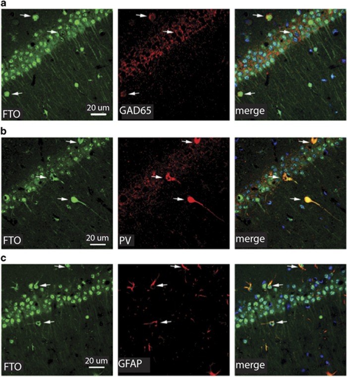
Figure 1.
Cellular and subcellular distribution of FTO in dorsal hippocampus. (a–c) FTO (green) is robustly expressed in CA1 neurons in the dorsal hippocampus. In neurons, this expression is robust in the cell body, but also observed in dendrites. (a) FTO is also expressed in glial cells and interneurons, as evidenced by co-expression of FTO with cells expressing the GABA cell marker GAD65 (red), (b) the interneuronal marker parvalbumin (PV, red), or (c) the glial marker GFAP (red). The merged image contains the DNA stain DAPI (blue). Arrows indicate the same cell across images.

