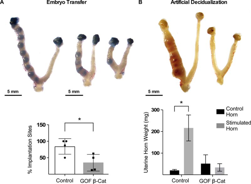
Figure 1. GOF β-catenin mice show reduced embryo implantation and decidualization.

(A) Embryos (E 3.5) were surgically transferred into one uterine horn of control (n=4) or GOF β-catenin (n=4) mice on pseudopregnancy day 2.5 and implantation sites were detected by Chicago blue dye. The number of implantation sites as a percentage of total embryos transferred per mouse were graphed and analyzed by unpaired T-test (P=0.03). (B) Control (n=3) and GOF β-catenin (n=3) mice were mated to vasectomized male mice to induce pseudopregnancy. On day of pseudopregnancy (DOPP) 4, oil was injected into one uterine horn to induce decidualization and uteri were collected on DOPP 9. Gross anatomy images show decidualization and was quantified by uterine wet weights. Values are the mean +/− s.e.m. compared by 2-way Anova, P=0.01. Representative images of uteri are shown.
