Figure 3.
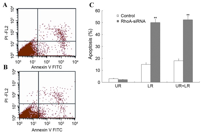
Level of apoptosis in cultured RhoA siRNA-transfected SPCA1 lung cancer cells. (A and B) RhoA siRNA-transfected cells and controls were collected and stained with annexin V (apoptosis stain) and PI (nuclear stain), and analyzed by flow cytometry. (C) The results from one of three independent assays are presented. The level of apoptosis in RhoA siRNA-transfected cells was significantly greater when compared with the control cells. **P<0.01 vs. control. RhoA, Ras homolog family member A; siRNA, small interfering RNA; UR, upper right; LR, lower right; PI, propidium iodide; FITC, fluorescein isothiocyanate.

