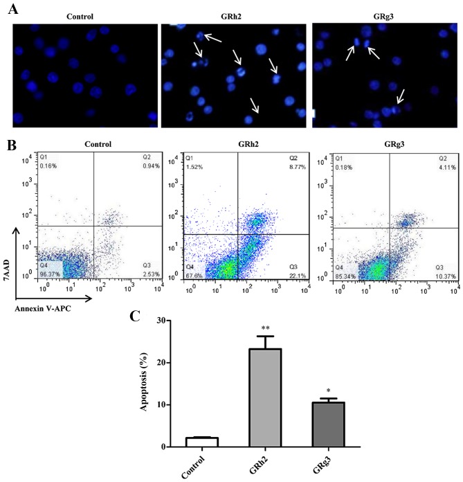
Figure 2.
GRh2 and GRg3 treatment induces apoptosis in Jurkat cells. Cells were treated with 35 µM GRh2 and GRg3 for 24 h. (A) Apoptotic cell death was examined with Hoechst staining by fluorescence microscopy (magnification, ×200). The white arrows indicate morphological alterations in the nuclei of Jurkat cells. (B) Annexin V-APC-positive and 7-AAD-negative stained cells indicate early apoptotic cells. The percentage of early apoptotic cells was analyzed by flow cytometer. (C) Quantification of the percentage of early apoptotic cells. Data are presented as the mean ± standard error of the mean (n=3/group) *P<0.05 and **P<0.01 vs. Control. GRh2, ginsenoside Rh2; GRg3, ginsenoside Rg3; APC, allophycocyanin; 7-AAD, 7-amino-actinomycin D.

