Figure 5.

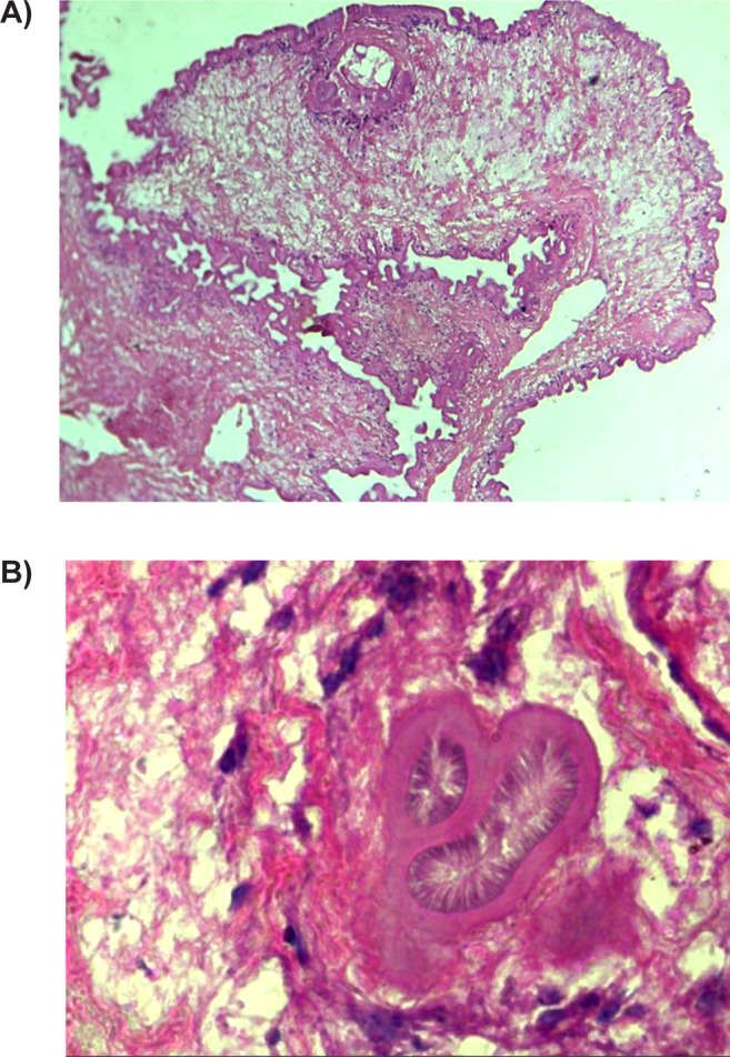
A) Photomicrograph (×200) in hematoxylin and eosin stain showed trilaminated corrugated cell wall structures with well defined protoscolex. B) Photomicrograph (×400) showed adjoining suckers in cysticercosis.

A) Photomicrograph (×200) in hematoxylin and eosin stain showed trilaminated corrugated cell wall structures with well defined protoscolex. B) Photomicrograph (×400) showed adjoining suckers in cysticercosis.