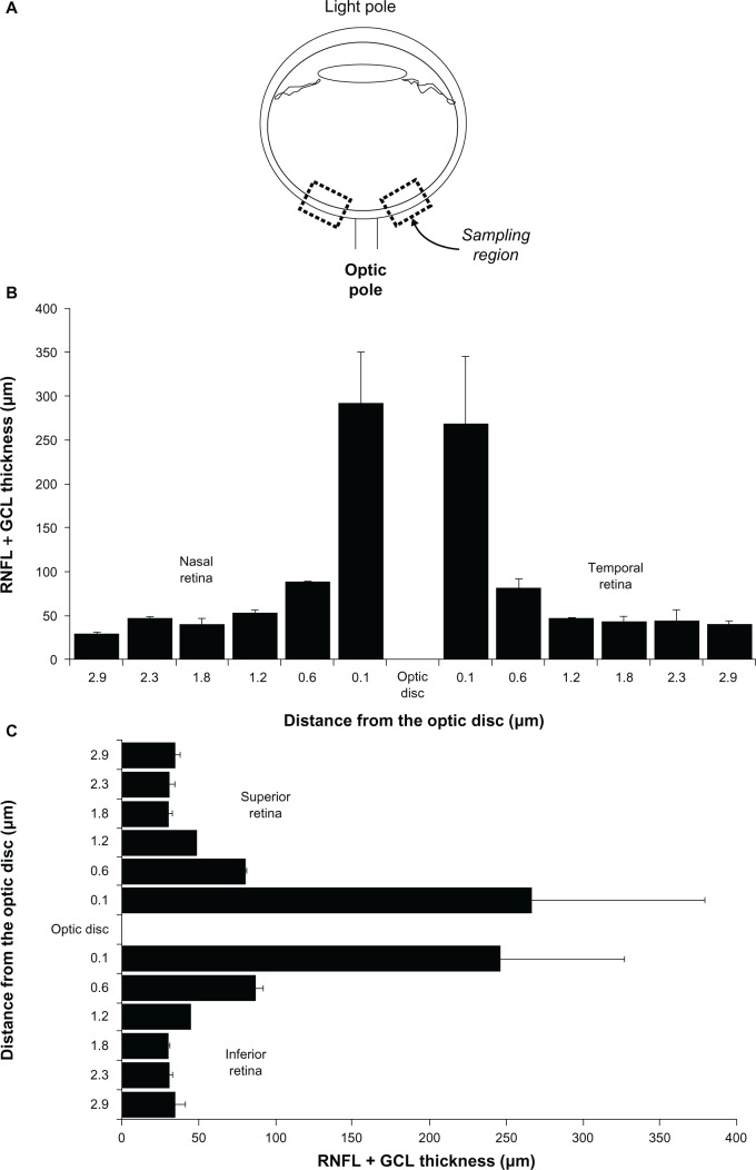
Figure 5.
Sampling strategy for retinal thickness, and retinal thickness per quadrant in the adult male rat. A) Samples of retinal images are systematically obtained from sagittal eyeball sections. These samples are located within 200 μm of the optic disc. In coronal eyeball sections B) and C) retinal thickness in the periphery decreases as a function of the distance to the optic disc, but there are no thickness differences between quadrants.

