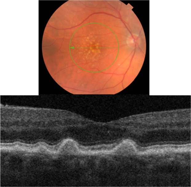
Figure 9.

Fundus photograph (top) showing hard cuticular drusen in the right eye of a patient with ARMD; the B-scan line on the fundus photograph has the same width as the B-scan SD-OCT image (bottom) which demonstrates the appearance of hard cuticular drusen.
Abbreviations: SD-OCT, spectral domain optical coherence tomography; ARMD, age-related macular degeneration.
