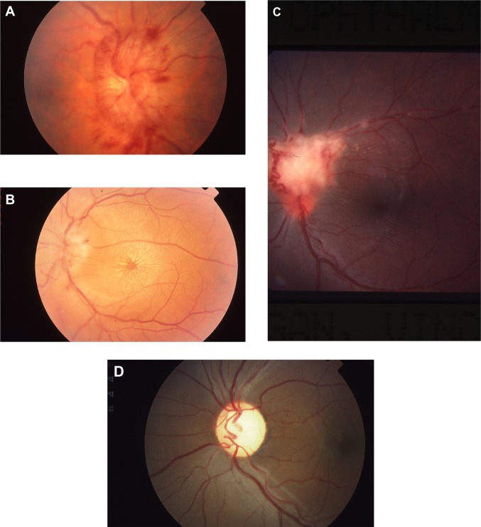
Figure 1.
Fundus findings in sarcoid optic neuropathy. A shows marked swelling with nerve fiber layer hemorrhages. The exudates in B form a star figure centered on the macula. C shows a mass on the disc consistent with granuloma and also shows sheathing of retinal vessels and optociliary shunt vessels. (D) Optic atrophy.

