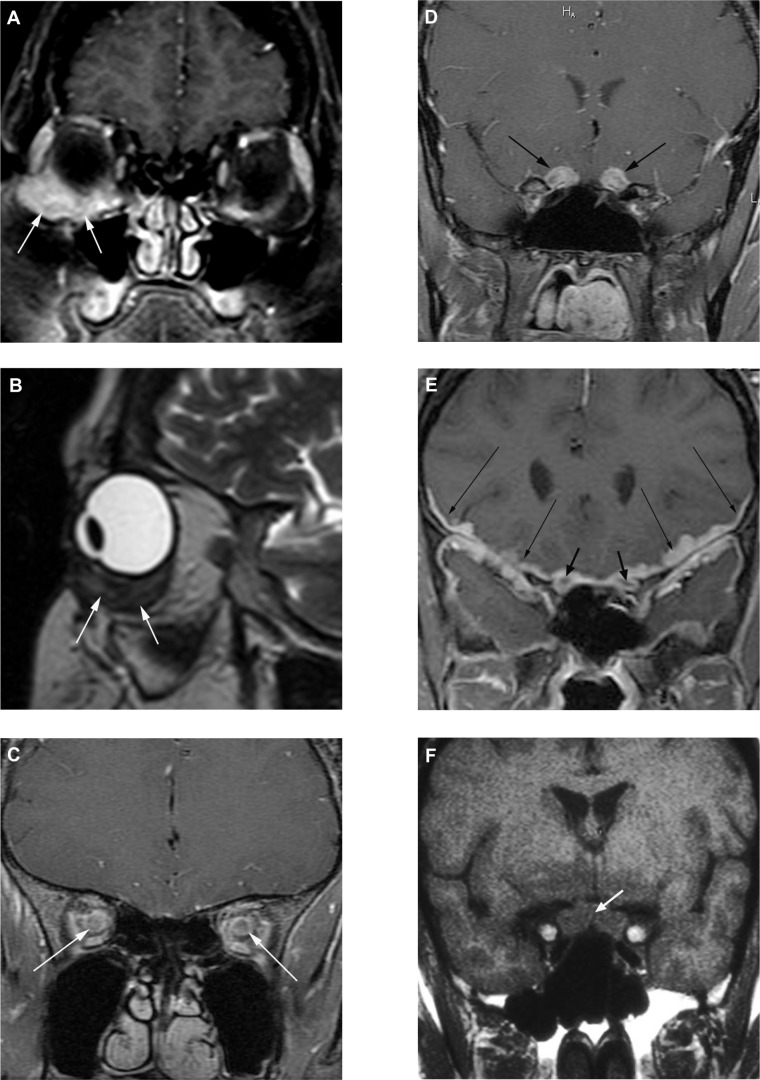
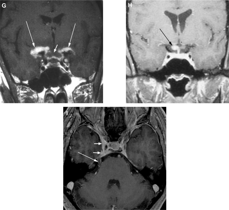
Figure 2.
Magnetic resonance imaging images of manifestations of neuro-ophthalamic sarcoidosis. (A) Coronal fat-suppressed T1-weighted post-contrast and (B) sagittal T2-weighted images demonstrate an ill-defined contrast-enhancing, low T2-signal inferior right orbital mass (arrows) surrounding the insertion of the inferior rectus muscle. Subsequent biopsy revealed granulomatous inflammatory tissue compatible with sarcoid. In another patient, coronal fat-suppressed T1-weighted post-contrast images reveal abnormal enhancement along the bilateral intraconal (C) and pre-chiasmal (D) optic nerves. In a third patient, (E) a coronal fat-suppressed T1-weighted post-contrast image reveals thick abnormal enhancement about the bilateral optic nerves (short arrows) and more extensively along the bilateral leptomeninges and dura (long arrows). In a fourth patient, presenting with blindness, a coronal pre-contrast T1-weighted image (F) and corresponding post-contrast image (G) demonstrate a swollen optic chiasm (short arrows) with thick leptomeningeal enhancement about and beyond the chiasm bilaterally (long arrows). In a fifth patient, a coronal fat-suppressed T1-weighted post-contrast image (H) reveals asymmetric right-sided involvement of the optic chiasm (arrow). In a sixth patient, presenting with right sided cranial nerves III and V abnormalities, an axial fat-suppressed T1-weighted post-contrast image (I) reveals involvement of the right cavernous sinus (short arrows) and along the cisternal portion of the ipsilateral trigeminal nerve (long arrow).


