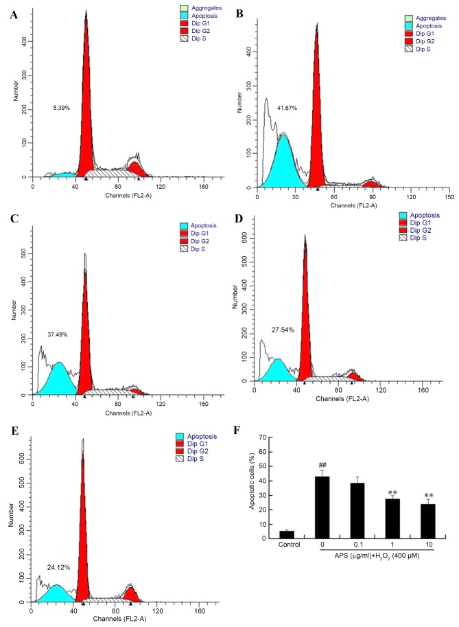
Figure 2.
Apoptosis of human umbilical vein endothelial cells, as determined by flow cytometry. (A) Control, (B) H2O2 only, (C) 0.1 µg/ml APS + H2O2, (D) 1 µg/ml APS + H2O2 and (E) 10 µg/ml APS + H2O2 groups. (F) The effect of APS on the percentage of apoptotic cells induced by 400 µM H2O2. Data are expressed as the mean ± standard deviation. ##P<0.01 vs. control; **P<0.01 vs. H2O2 only. APS, Astragalus polysaccharide.

