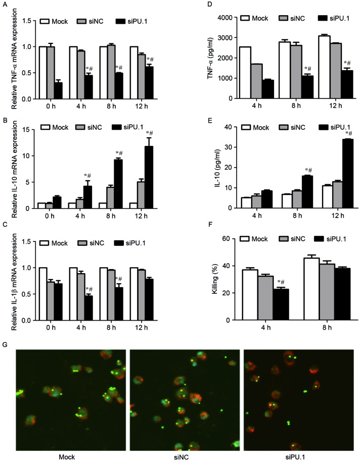
Figure 4.
mRNA levels of (A) TNF-α, (B) IL-10 and (C) IL-1β in mock, siNC, or siPU.1-transfected THP-1 cells at 0, 4, 8 and 12 h of exposure to resting conidia, as determined by reverse transcription-quantitative polymerase chain reaction analysis. The protein levels of (D) TNF-α and (E) IL-10 were assayed by enzyme-linked immunosorbent assay analysis. (F) Killing ability was assessed by serial dilution of cells following co-incubation with resting conidia, and quantification of the number of colony forming units. (G) Confocal laser-scanning microscopy was used to observe phagocytosis after 4 h of incubation with resting conidia. Magnification, ×63 (objective oil immersion lens). *P<0.05 vs. Mock group; #P<0.05 vs. siNC group. TNF, tumor necrosis factor; IL, interleukin; Mock, untransfected cells; siNC, negative control small-interfering RNA; siPU.1, small-interfering RNA targeting PU.1.

