Figure 1.
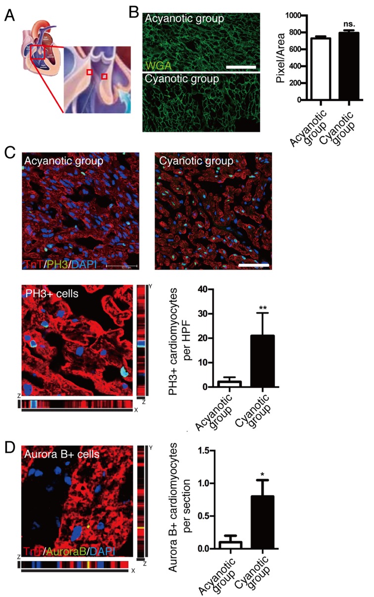
Effect of hypoxia on cardiomyocyte proliferation in human infants. (A) Myocardial tissues were taken from the distal obstructive right ventricular outflow tract. (B) Cardiomyocyte cell size was not significantly different between acyanotic and cyanotic infants; scale bar, 50 µm. (C) Coimmunostaining with pH3 and cardiac TnT antibodies demonstrated a significant increase in cardiomyocyte mitosis in the myocardium of cyanotic patients compared with acyanotic patients; scale bar, 50 µm. (D) Representative image of coimmunostaining with anti-Aurora B and cardiac TnT antibodies demonstrated increased cytokinesis in the myocardium of cyanotic patients. Data is presented as the mean + standard error of the mean. *P<0.05 and **P<0.01 vs. acyanotic group. PH3, phospho histone H3 Ser10; TnT, troponin T; WGA, wheat germ agglutinin; HPF, high power field; P, days after birth; ns., not significant.

