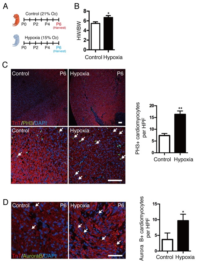
Figure 2.
Effect of hypoxia on cardiomyocyte proliferation in neonatal mice. (A) Neonates were exposed to a normoxic or hypoxic environment at birth for 6 days. (B) HW/BW ratio was significantly increased in mice exposed to hypoxia. (C) Coimmunostaining demonstrated an increase in cardiomyocyte mitosis in hypoxia-exposed hearts; scale bar, 100 µm. (D) Coimmunostaining with anti-Aurora B and anti-TnT antibodies demonstrated increased cytokinesis in hypoxic hearts; scale bar, 50 µm. Data is presented as the mean + standard error of the mean. *P<0.05 and **P<0.01 vs. control. HW/BW, heart weight/body weight; TnT, troponin T; P, days after birth; PH3, phospho histone H3 Ser10; HPF, high power field.

