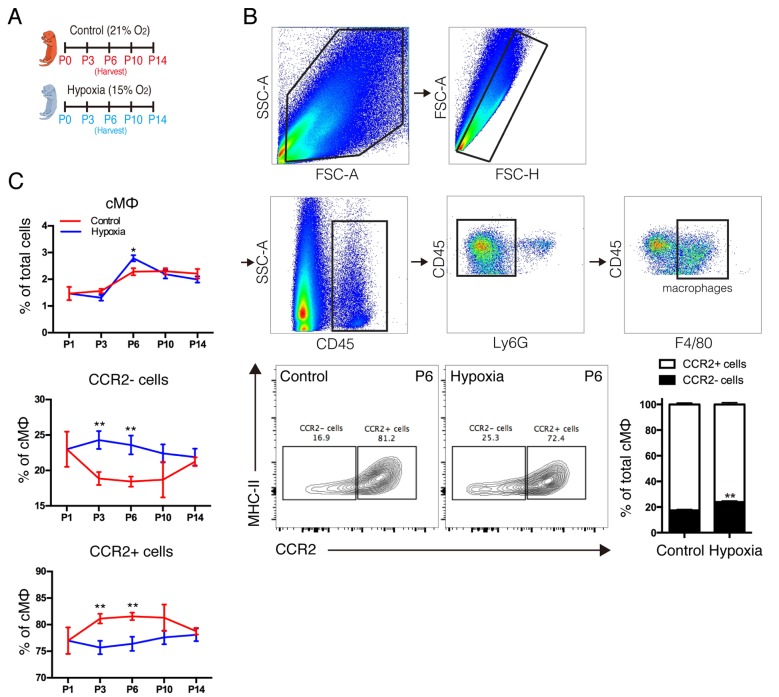
Figure 3.
Effect of hypoxia on distinct lineages of cardiac macrophages. (A) Neonates were exposed to a normoxic or hypoxic environment at birth for 2 weeks. (B) Gating strategy and representative fluorescence activated cell sorting analysis of macrophage populations in the neonatal heart on 6th day under normoxic and hypoxic conditions. (C) Quantification of macrophage populations in the neonatal heart in normoxic and hypoxic environments at various time points after birth. Data is presented as the mean ± standard error of the mean. *P<0.05 and **P<0.01 vs. control. P, days after birth; SSC, side scatter; FSC, forward scatter; Ly6G, lymphocyte antigen 6 complex locus G; CCR2, C-C chemokine receptor type 2; MHC II, histocompatibility-2 MHC; cMΦ, cardiac macrophages.

