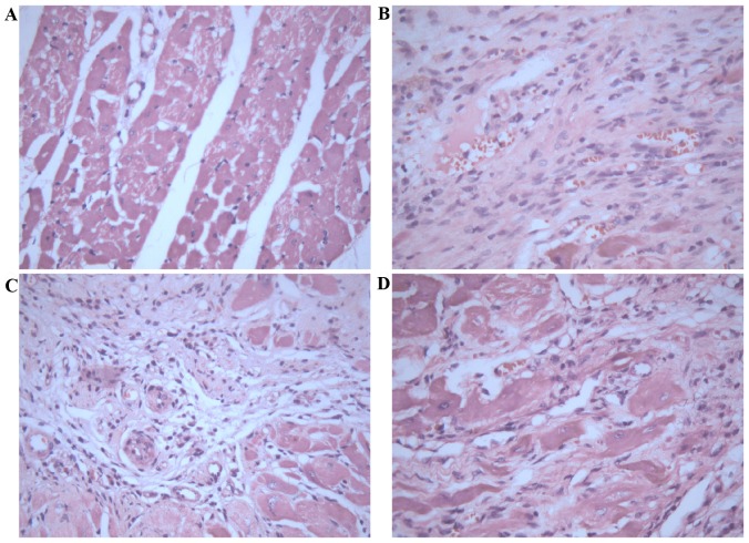
Figure 2.
Morphological analysis of myocardial tissue stained with hematoxylin-eosin. (A) Rats in the sham control group exhibited apparent integrity of the myocardial cell membrane, with bright red cytoplasms and oval nuclei in the center of cells. (B) Rats in the myocardial infarction model group exhibited an irregular arrangement of myocardial cells, dissolved nuclei, a large number of fibroblasts or fibers, and ruptured of myocardial fibers. Treatment with (C) 2.5 and (D) 10 mg/kg/day Astragaloside attenuated these histopathological alterations. Magnification, ×400.

