
Figure 5. C5a expression and binding in the inflamed joint.
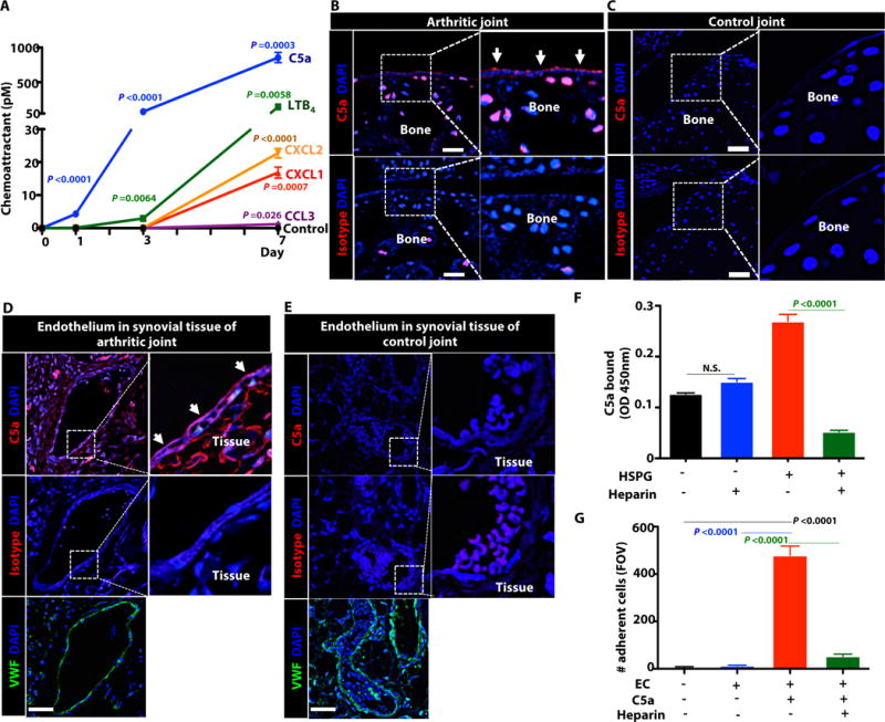
(A) Levels of CAs in SF on days 0, 1, 3 and 7 following AST as determined by ELISA. n = 3 mice/time point (B–E) Immunofluorescence staining of joint tissue from arthritic mice on day 7 after AST (B, D) or control mice (C, E). Red: C5a; Green: VWF+ECs; Blue: DAPI. Scale bars =50μm. Data are representative of 3 independent experiments. (F) In vitro binding assay. Amount of C5a bound to uncoated 96well assay plates or plates coated with HSPG (5μg/ml) in the presence or absence of heparin (80μg/ml) (G) In vitro adhesion assay. Number of BM neutrophils adherent to murine aortic endothelial cells that were pretreated with C5a (1nM) in the presence or absence of heparin (80μg/ml) (F,G) n = 3 independent experiments. Data indicate mean ± SEM; P value calculated using unpaired two-tails Student’s t-test. N.S.- not statistically significant.