
Figure 7. β2 integrin activation mediates C5a-induced neutrophil adhesion to the joint endothelium.
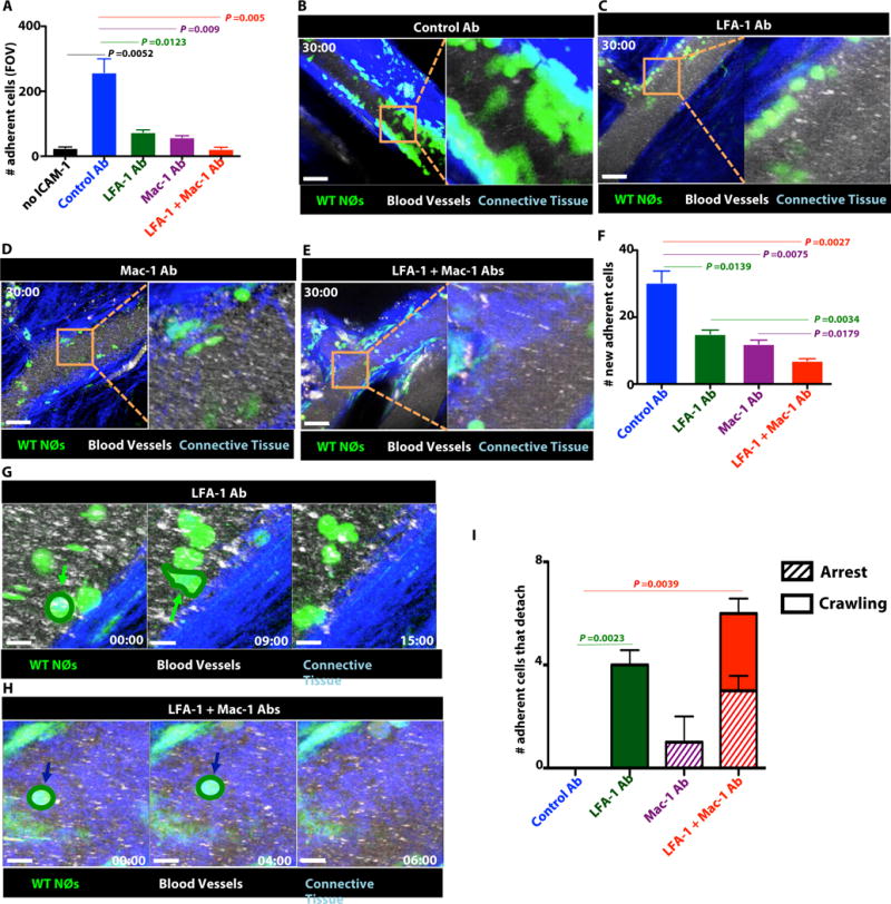
(A) In vitro adhesion of C5a (1 nM) activated WT neutrophils to ICAM-1 coated plates in presence of β2 integrin blocking mAbs or an isotype control mAb. n = 3 independent experiments. Data indicate mean ± SEM; P value calculated using unpaired two-tails Student’s t-test. (B–F) In vivo imaging of joints in WT-LysM-GFP mice that were pretreated for 4 hrs with β2 integrin blocking mAbs or an isotype control mAb following exposure to C5a (1nM). (B) Control Ab, (C) LFA-1 Ab, (D) Mac-1 Ab (E) LFA-1 and Mac-1 Ab treated mice. Green: neutrophils; Blue: Connective tissue; Qdots: Blood vessels. Scale bars represent 50 μm. Time in mins:secs. Data are representative of 3 independent experiments. (F) Number of newly adherent cells on the joint endothelium over the 30 mins of observation. (G–I) Neutrophil detachment in vivo. (G) LFA-1 Ab, (H) Both LFA-1 and Mac-1 Ab treated mice. Green: neutrophils; Blue: Connective tissue; Qdots: Blood vessels. Green arrows indicate crawling cells that detached and blue arrows indicate arrested cells that detach. Time in mins:secs. Scale bars represent 10 μm. Data are representative of 3 independent experiments. (I) Number of newly adherent cells that detach over the 30 mins of observation. Striped bar indicates arrested cells that detach and closed bar indicates crawling cells that detach of the total number of adherent cells that detach. (F, I) n = 3 mice/group. Data indicate mean ± SEM; P value calculated using unpaired two-tails Student’s t-test.