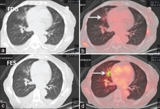
Figure 2.

Lung window computed tomography and fused 18F-fluorodeoxyglucose (a and b) and 18F-fluoroestradiol (c and d) positron emission tomography-computed tomography axial images: 18F-fluorodeoxyglucose avid prominent bronchial markings with peribronchial infiltrates in the right middle lobe which shows good 18F-fluoroestradiol uptake (white arrows). Findings favor lymphangitis carcinomatosis
