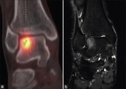
Figure 2.

Lateral talar dome osteochondral lesion in a 32-year-old male. (a) Coronal fused technetium-99m methylene diphosphonate single-photon emission computed tomography/computed tomography image demonstrates a subchondral lucency in keeping with Type 5 lesion as described by Loomer et al. with surrounding increased uptake of tracer. The subchondral bone plate appears intact. (b) Coronal short tau inversion recovery image demonstrates a subchondral abnormality which correlates with the site of the lucency seen on the corresponding single-photon emission computed tomography/computed tomography image. Note the discrepancy between the area of increased uptake on (a) single-photon emission computed tomography/computed tomography and high signal marrow edema on the (b) magnetic resonance imaging
