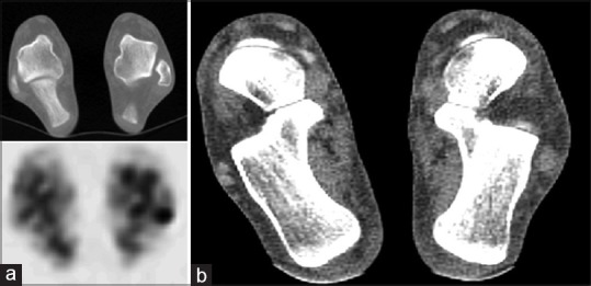
Figure 6.

Left peroneal tendinosis in a 48-year-old male. (a) Axial unfused technetium-99m methylene diphosphonate single-photon emission computed tomography/computed tomography images demonstrate increased uptake at the posterior aspect of the left lateral malleolus which demonstrates no associated bony abnormality on the computed tomography component of the study. (b) Axial image from the computed tomography component (soft tissue windows) at a more inferior slice demonstrates thickening and poor definition of the left peroneal tendons when compared to the normal right side
