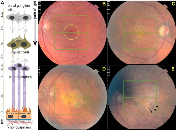
Figure 1.
Structure of the retina and development of age-related macular degeneration (AMD) pathology.
(A) Schematic diagram depicting cross-section of the retina and associated tissues. Retinal ganglion cells (RGCs), inner plexiform layer (IPL), inner nuclear layer (INL), outer plexiform layer (OPL), outer nuclear layer (ONL), photoreceptor inner segments (IS), outer segments (OS), retinal pigment epithelium (RPE), Bruch's membrane (BrM) and choriocapillaris. Layers are shown juxtaposed to specific cell-types. (B) Funduscopy image of a representative healthy retina as viewed through an ophthalmoscope. The macula is denoted by a yellow circle. (C) The appearance of macular drusen (yellow-white spots) is considered to be the first clinical indicator of AMD. (D) Disease may progress to geographic atrophy (GA) in which RPE and overlying photoreceptors are obliterated resulting in a macular lesion, or (E) vascular AMD in which invasive/leaky choroidal vessels cause a haemorrhage (black arrows) to result in retinal damage.

