Figure 1.
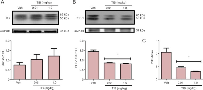
Changes in the content of total Tau and PHF-1 in the hippocampus of aged mice treated with tibolone (TIB).
(A) Western blots for total Tau content in the hippocampus of aged mice. (B) PHF-1 levels in the hippocampus of aged mice treated with TIB. GAPDH was used to correct differences of total loaded protein. Proteins detected by western blot analysis from the hippocampus of mice treated with TIB were quantified by densitometric analysis and corrected using GAPDH protein content data. (C) The ratio of PHF-1 and Tau to their respective control (GAPDH) expression in the hippocampus of mice treated with low- and high-dose TIB. Results are expressed as the mean ± SE of six individual experiments. *P < 0.05, vs. vehicle (Veh).

