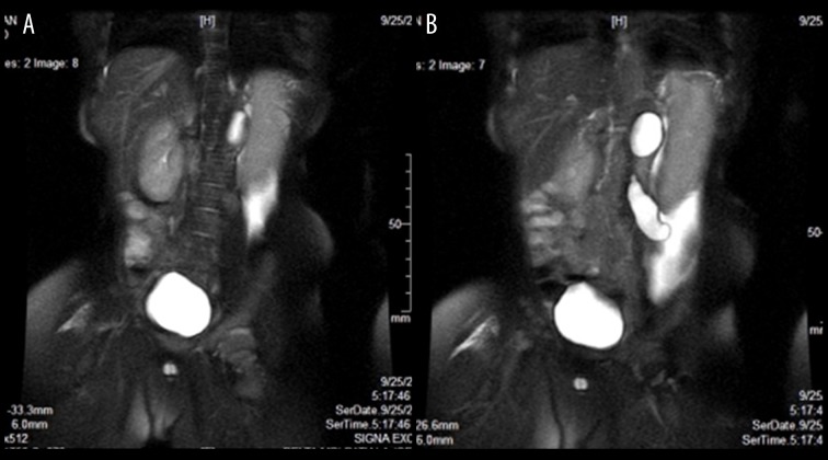
Figure 5.
(A) MRI abdomen, T2 W, coronal view, shows right kidney is enlarged, but normal in morphology, the left kidney shows cystic dysplasia, ascites is seen in left lower lumbar region. (B) MRI abdomen, T2 W, coronal view, shows right kidney is enlarged, but normal in morphology, the left kidney shows cystic dysplasia, left ureter is dilated and tortuous, (the tubular cystic structure which was seen on ultrasound in the left lumbar region), ascites is seen in left lumbar region.

