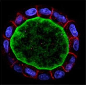
Fig. 1.

Confocal section of a Madin-Darby canine kidney (MDCK) cyst grown in Matrigel. Cells form a spherical cyst in the first step of renal tubulogenesis (apical membrane and lumen: green; nucleus: blue; basolateral membrane: red; staining and related information on cyst and markers used can be obtained from [97]). Photo courtesy of Sang-Ho Kwon and Keith Mostov
