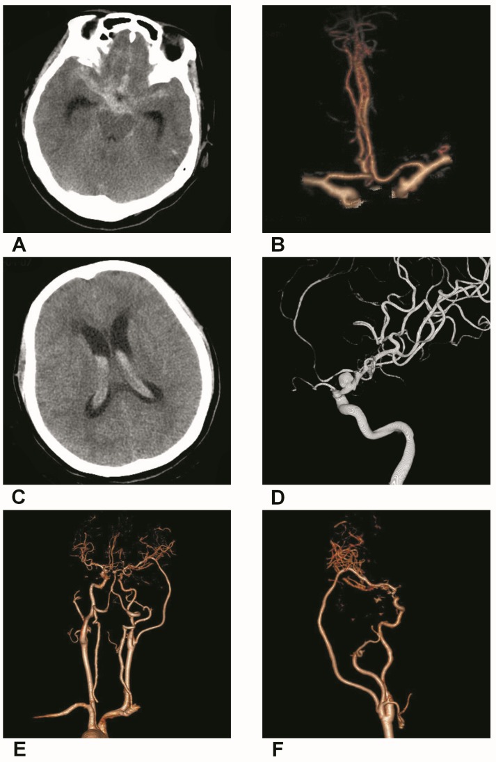
Figure 3.
Trapping with bypass of BBAs of the supraclinoid ICA. A, CT of the first SAH showing the hemorrhage in the suprasellar cistern. B, CTA showing a small BBA of the supraclinoid ICA. C, CT of the second SAH showing the intraventrical hemorrhage. D, DSA showing the BBA became bigger. E-F, Postoperative CTA showing that the BBA was trapped with EC-middle artery bypass.

