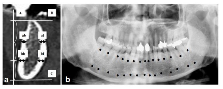
Figure 1.
Method to measure the cortical bone width in mandible using Dental CT, originally established by Hamada et al. [8]: (a) = Measurement sites in oblique sagittal images. A line was drawn vertical to the occlusal plane [A], and 2 lines were marked parallel to the occlusal plane which divided the space between the alveolar crest [B] and the lower edge of the mandible [C] into 3 equal parts. ↔: Cortical bone width was defined as [ab + al] on the alveolar level and [bb + bl] on the body level. ab: alveolar buccal, al: alveolar lingual, bb: body buccal, bl: body lingual. (b) = Measurement points in each patient. A total of 15 sites was between the teeth from the second molar of one side to the second molar of the opposite side. Measurements were made at 30 points [●] in the alveolar border and the mandibular body, resulting in 15 oblique sagittal images of DentaScan. [ab + al + bb + bl] ÷ 2 was calculated at 15 points. The average of 15 points in each case was defined as the value of cortical bone width and used for comparison.

