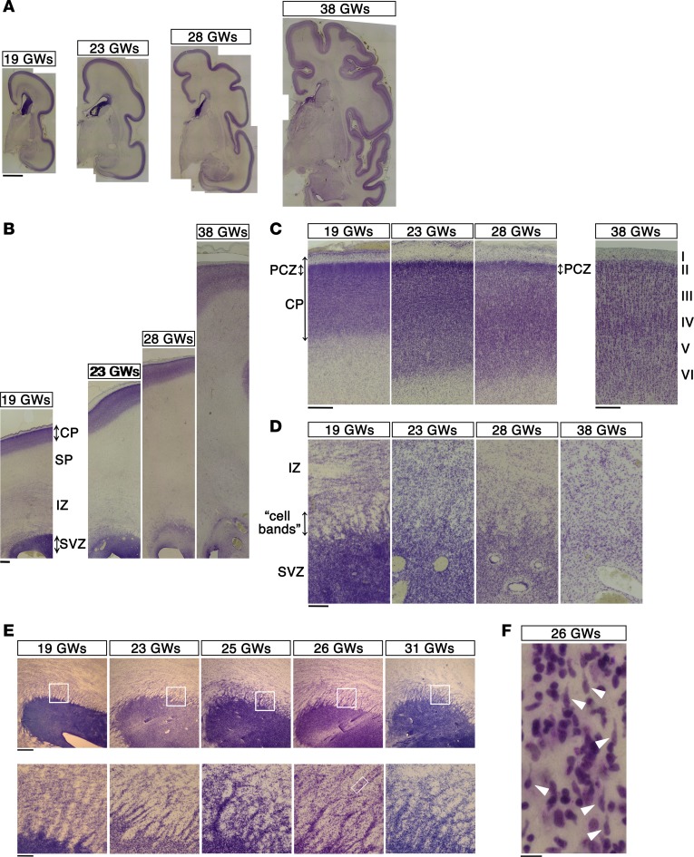
Figure 1. Histological features of the developing human neocortex.
(A) Coronal sections from the human neocortex at 19, 23, 28, and 38 GWs were stained with Cresyl violet (Nissl staining). Scale bar: 5 mm. (B) High-magnification images of the dorsolateral neocortex in A. Scale bar: 1 mm. SP, subplate.(C) High-magnification images of the forming cortical plate (CP) in B. Note that the outermost regions of the neocortex at 19, 23, and 28 GWs, but not at 38 GWs, showed cell-dense accumulation of neurons. This region is thought to correspond to the primitive cortical zone (PCZ). Scale bar: 500 μm. (D) High-magnification images of the subventricular zone (SVZ) in B. Numerous “cell bands” radiating from the SVZ toward the intermediate zone (IZ) were recognized until 28 GWs but became faint by 38 GWs. Scale bar: 200 μm. (E) The “cell bands” were particularly prominent in horizontally sliced sections of the neocortex at 19 to 31 GWs (lower-magnification images are shown in Supplemental Figure 1). Boxed areas in the top panels are shown at high magnification in the bottom panels. Scale bar: 500 μm (top); 100 μm (bottom). (F) A further magnification of the boxed area in the bottom panel in E (26 GWs). The “cell bands” contained plenty of cells with the morphological features of migrating neurons (white arrowheads). Seven focal planes were merged to visualize the shapes of the entire cells. Scale bar: 10 μm.

